Purpose of mastoid x-ray
This is an automatically translated article.
X-ray of the mastoid bone is one of the imaging tools that is very useful in diagnosing inflammatory and infectious diseases in the mastoid bone. In addition, a mastoid X-ray may also be ordered in cases of trauma. This article will provide some essential information about mastoid X-ray
1. What is the mastoid bone of the body?
Mastoid is a small bone in the skull. The mastoid bone is located just behind the auricle, protrudes outward and is palpable. In the anatomical position, the mastoid is located below, posteriorly, and outward relative to the temporal bone. Adjacent to the mastoid bone are many important organs such as the brain, meninges, blood vessels and several important nerves. Therefore, when there is a disease that damages the mastoid ear, it can easily spread to other organs. The mastoid bone has an extremely important function for the body's hearing function. Although considered a bone of the skull, the mastoid is not intrinsically a typical skeletal structure. Inside the mastoid bone there are many sponge-like air sacs, which are different from the dense and rough structure found in other bones of the body. These air-filled structures protect the ear's villi, regulate pressure in the ear, and help protect part of the temporal bone if an injury occurs.

2.Some diseases in mastoid region
There are two common diseases of the mastoid bone, which often stem from inflammatory and infectious foci in the ear such as otitis media. If left untreated, it can easily lead to mastoid otitis.
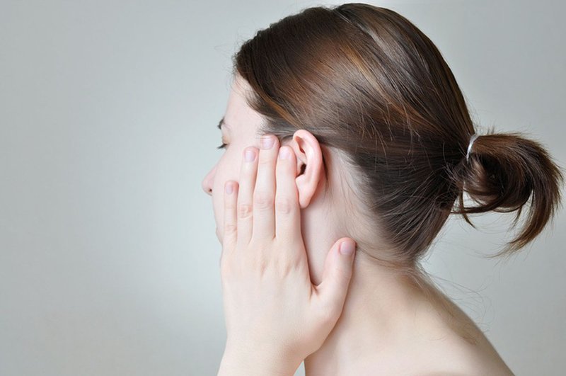
When mastoiditis is present, the patient may experience dizziness, tinnitus, hearing loss. If it is more severe, it can lead to inflammation and infection of surrounding important organs such as encephalitis - meningoencephalitis, epidural abscess, papilledema or sepsis.
To diagnose mastoiditis, x-ray of mastoid is a commonly used tool in clinical practice.
3. What is mastoid x-ray?
Mastoid X-ray, also known as Schuller X-ray. This X-ray technique is taken in the temporomandibular position. This is the most common position and is often used when imaging technicians take pictures of patients.
On the mastoid X-ray film (Schiller's position), mastoiditis images can see the hollows in the mastoid bone, which are thick and opaque due to the destruction of cell groups in the mastoid bone, there are lesions that have lost their wall and become widespread inflammation.
Additionally, a mastoid x-ray may be indicated in some head injuries involving the mastoid.

4. General techniques of Schuller X-ray:
In case the patient has signs of ear pain or pain due to an ear infection or due to many different causes causing pain in the ear. The radiologist or radiologist may order Schuller X-rays.
To take an X-ray of the mastoid, the patient needs to have:
The Schuller X-ray position helps the image on the film to reveal the two sides of the striatum, the atrial septum and the temporomandibular joint. From there, the doctor can clearly see and evaluate the lesions located in the mastoid sinus and temporomandibular joint. The technician will guide the patient to lie on his side from the lateral cephalometric position. The main X-ray source is located at 25-300 degrees from the axis of the two ears. Or the center of the source can be adjusted up to about 7cm from the ear canal on the opposite side and the penetrating ray can pass through the ear canal on the shooting side. In addition, the auricle of the right side folds forward so that the image does not cover the mastoid bone. The imaging criteria for the Schuller X-ray technique are as follows:
The outer and inner ear canals overlap and must be at the level of the temporomandibular joint. The ear flaps fold forward. Acute and chronic otitis media. Radiology technician preparation steps for X-ray Schuller
Prepare X-ray machine (DR or CR system) Film printer, storage system. HIS network system and automatic number calling. For patients who come into contact with imaging devices, it is necessary to remove jewelry and metal objects, if any.

The patient is lying prone on the imaging table, the right hand to be taken is placed down on the body, the opposite hand is placed in the position right in front of him. Gradually adjust the patient's position to lie on the side, the plane of the head is parallel to the film, the patient's head is slightly bent so that the Virchow plane is parallel to the upper border of the film, the lateral atrium near the film is in the center of the film. Adjust the lamp head towards the foot 25-300, select a point 5cm above and 5cm behind the outer ear canal distal to the film, directing the beam through the atrium to be imaged. Observe the patient through the lead glass of the control room, press the X-ray generator button See the satisfactory technical image. Instruct the patient to leave the shooting position. Technical processing of images on computers and film printing.
5. Purpose of mastoid X-ray technique
Temporomandibular joint disease The ear does not develop. Cholesteatoma in the ear (dead bone surrounded by fatty tissue). Suspected fracture of the stone bone. Semicircular fistula.

Processing of results after Schuller X-ray
Normal: clearly visible cysts and their septum.
Pathological case:
Blurred septum, indistinct septum in acute mastoiditis. Osteoarthritis, septum loss in chronic mastoiditis. On the background of mastoid is blurred, there is a bright area, surrounded by a bold border, floating like a cloud, thinking of cholesteatoma lesions in chronic mastoiditis with cholesteatoma. X-ray techniques as well as many other medical examination and treatment techniques at Vinmec International General Hospital are conducted by a team of highly qualified and experienced medical doctors; the system of advanced and modern machinery should significantly reduce radiation dose with the best image and always be updated according to medical progress in the world; Especially, professional service quality will help customers have the most comfortable and secure experience when visiting Vinmec.
Please dial HOTLINE for more information or register for an appointment HERE. Download MyVinmec app to make appointments faster and to manage your bookings easily.
-
 Uses of Fonclar
Thuốc Fonclar được dùng theo đơn của bác sĩ để đẩy lùi hiệu quả các tình trạng nhiễm khuẩn như viêm phế quản, viêm phổi, viêm amidan,... Bệnh nhân cần tham khảo kỹ lời khuyên của bác sĩ về cách ...
Readmore -
 Medicine for acute mastoid otitis
Viêm tai xương chũm cấp tính là tình trạng bệnh có thể gặp ở nhiều đối tượng khác nhau, bệnh được đánh giá là khá nguy hiểm nếu không điều trị kịp thời. Vậy chữa viêm tai xương chũm cấp ...
Readmore -
 Uses of Mulasmin
Thuốc Mulasmin 500 là thuốc kháng sinh đường uống có thành phần chính là Azithromycin 500mg. Cũng như các loại kháng sinh thông thường, chỉ dùng thuốc Mulasmin theo chỉ định của bác sĩ cho những trường hợp nhiễm khuẩn ...
Readmore -
 Uses of Coducefa
Coducefa là một loại kháng sinh đường uống dùng theo đơn, người bệnh tuyệt đối không tự ý sử dụng khi chưa có chỉ định. Cùng tìm hiểu rõ hơn thuốc Coducefa có công dụng gì? Liều dùng Coducefa thế ...
Readmore -
 Uses of Cefvalis
Cefvalis là kháng sinh dùng theo đơn. Tuân thủ chỉ định, liều dùng thuốc Cefvalis sẽ giúp người bệnh nâng cao hiệu quả điều trị và tránh được những tác dụng phụ không mong muốn.
Readmore