
This article has been professionally consulted by Dr. Nguyen Thanh Hai, Grade I Specialist, a specialist in Diagnostic Imaging - Department of Diagnostic Imaging and Nuclear Medicine - Vinmec Times City International Hospital. Dr. Hai has over 20 years of experience in the field of diagnostic imaging, particularly in multidetector computed tomography and magnetic resonance imaging.
A herniated disc is a condition wherein the nucleus pulposus protrudes from the outer fibrous layer of the disc, subsequently compressing adjacent structures and causing pain in patients. Therefore, X-rays are unable to detect the condition in its early stages; however, when the nucleus pulposus has extensively extruded, compressing nerve roots and encroaching upon the spinal cord, X-rays may assist in the diagnosis.
1. What is a herniated disc?
A herniated disc is a condition that, while not directly life-threatening, can significantly impair a patient's mobility and overall health.
The primary cause of a herniated disc is the displacement of the nucleus pulposus from its original position, leading to compression of nerve roots and prolonged discomfort for the patient. Additional risk factors for herniated discs include:
• Age: The aging process leads to dehydration and degeneration of the discs and spine, making them more susceptible to injury, resulting in herniated discs.
• Back injuries, overexertion, and incorrect postures can also contribute to damage to the discs and spine.
• Congenital spinal abnormalities or genetic predispositions.
• Obesity: Increased body weight places additional pressure on the spinal discs, particularly in the lumbar region, contributing to herniation.
If a herniated disc is not treated promptly, it can lead to severe complications such as nerve root compression, spinal canal narrowing that may impede mobility, loss of bowel control due to lumbar nerve root involvement, muscle weakness and atrophy from prolonged inactivity, and dysfunction of the urinary sphincter leading to urinary retention or incontinence.

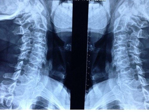
2. Can X-rays detect a herniated disc?
X-ray imaging is a method that employs external radiation to visualize internal changes in bones, joints, and other structures. The X-ray machine emits high-energy X-ray beams, which penetrate soft tissues and bodily fluids to produce images. Dense structures like bone absorb more radiation, reducing the penetration of X-rays.
So, can X-rays detect a herniated disc? This condition arises from the protrusion of the nucleus pulposus beyond the fibrous layer, compressing surrounding structures and causing pain. Consequently, X-rays cannot detect the condition in its early stages, as the spinal structure is minimally affected.
What is the purpose of X-ray imaging in the context of herniated discs? The utility of X-rays is limited to:
• Diagnosis in cases where the nucleus pulposus has significantly extruded, compressing nerve roots, producing spinal deformities, or narrowing the intervertebral spaces.
• Furthermore, X-ray images can provide insights into the current alignment of the patient's spine, allowing for preliminary assessments of early signs related to lumbar and cervical disc degeneration.
• When a contrast agent is injected, radiographs of herniated disc can indirectly visualize the extrusion of the nucleus pulposus and identify any nerve root or spinal cord compression.
• This diagnostic approach may be optimal for patients with limited financial resources or those residing far from advanced medical facilities, as the obtained images can facilitate provisional diagnoses.
3. When should X-rays be conducted for herniated discs?
X-rays should be performed when patients exhibit the following common symptoms of herniated discs:
• Muscle weakness or paralysis: These symptoms typically emerge after prolonged lack of treatment, leading to disease progression. Mobility becomes increasingly difficult and may result in muscle atrophy and paralysis.
• Limb pain: Sudden onset of pain in the cervical or lumbar regions that radiates to the shoulders, arms, or legs warrants an X-ray. Pain may be persistent or acute during movement or physical exertion.
• Numbness in the limbs: The displacement of the nucleus pulposus outward may compress adjacent nerves, resulting in numbness and pain in the cervical and lumbar regions, which may spread to the buttocks, groin, thighs, and heels. Patients may experience altered sensation, often described as a "pins and needles" sensation.
• In addition, if the severity of pain, numbness, or muscle weakness intensifies and detrimentally impacts daily life, or if symptoms like urinary retention or incontinence or loss of sensation in the inner thigh, calf, and perianal region manifest, an X-ray should be promptly undertaken.
4. Procedure and Considerations for X-ray Imaging of Herniated Discs
4.1. Procedure for X-ray Imaging of Herniated Discs
Typically, the process of X-ray imaging for herniated discs is not overly complex. The steps are as follows:
• Step 1: Instruct the patient to remove any jewelry or clothing with metallic fastenings to avoid interference with the X-ray imaging process.
• Step 2: Request that the patient either stand upright or lie in a lateral position to facilitate the X-ray imaging of the herniated disc. The imaging process usually takes approximately 5 to 10 minutes, after which the radiologist will analyze the images, document findings, and provide results.
• Step 3: Upon receiving the X-ray films, if any abnormalities in the spine are observed, the patient should undergo additional examinations such as computed tomography (CT) or magnetic resonance imaging (MRI) to achieve the most accurate results.
4.2. Considerations for X-ray Imaging of Herniated Discs
To ensure that the X-ray imaging procedure is conducted safely and accurately, patients should be aware of the following points:
• Pregnant women should not undergo X-ray imaging for herniated discs as it may adversely affect fetal development.
• Remove any objects or devices such as hairpins and jewelry, as they may impact the results of the X-ray.
Patients are advised to visit hospitals with orthopedic specialties to undergo the X-ray imaging procedure, aiding in the timely detection of conditions and facilitating prompt treatment for herniated discs.

To arrange an appointment, please call HOTLINE or make your reservation directly HERE. You may also download the MyVinmec app to schedule appointments faster and manage your reservations more conveniently.