This article was written by Tran Thi Vuong, MSc, MD Microbiologist, Laboratory Department, Vinmec Hai Phong International General Hospital.
Gonorrhea is one of the most common sexually transmitted infections today. However, its symptoms are often easily mistaken for those of other diseases. Therefore, the stain and microscopy technique for detecting gonorrhea is highly valuable in diagnosis. How is this technique performed?
1. What is gonorrhea?
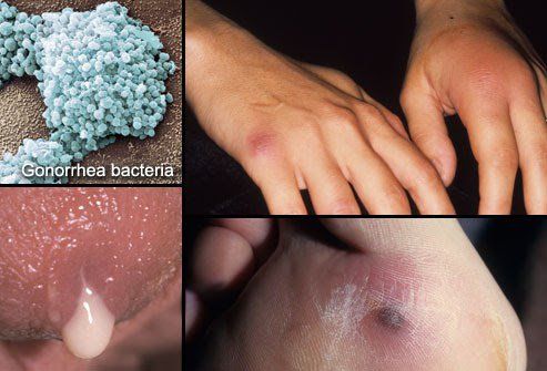
Gonorrhea is caused by Neisseria gonorrhoeae, which can be found in the vagina, cervix, eyes, mouth, anus, and male urethra. Gonorrhea can affect individuals of all ages but is most common among men and women of reproductive age. In women, gonorrhea can cause severe complications, including fallopian tube inflammation, ectopic pregnancy, miscarriage, or transmission from mother to child. Gonorrhea is mainly transmitted through sexual contact, but gonococcal conjunctivitis may occur in newborns infected during childbirth.
The symptoms of gonorrhea are often unclear, making it hard to identify and easy to confuse with other urinary tract infections. Approximately 10-20 days after being infected with the gonorrhea bacterium, patients begin to show symptoms. Specific symptoms of gonorrhea include:
- In men: Painful urination, burning sensation while urinating, frequent urination, and urine that may contain blood or pus. Severe cases may involve the discharge of pus resembling "banana resin" from the urethral opening, particularly in the morning, accompanied by fatigue and mild fever.
- In women: Symptoms are usually unclear and often overlooked or mistaken for common gynecological diseases. In advanced stages, women may experience painful urination, green or yellow pus discharge from the urethra or cervix, and vaginal odor.
2. The value of stain and microscopy in detecting gonorrhea
- Stain and microscopy involves staining urogenital specimens and observing them under a microscope to assess the morphology, color, and presence of specific bacteria or cells. For gonorrhea, this test evaluates the morphology, size, staining properties, and characteristic arrangement of Neisseria gonorrhoeae, as well as other cellular features.
- This technique is simple and commonly used for diagnosing genital infections, including gonorrhea. Its advantages include an easy-to-perform procedure, quick time, and reliable results, helping physicians determine appropriate treatment strategies.
- Besides detecting Neisseria gonorrhoeae, this test is also used to diagnose other diseases such as vaginosis, urethritis caused by bacteria, fungi or Trichomonas vaginalis,...
3. Indications for stain and microscopy for gonorrhea
This test is indicated in cases of suspected gonorrhea or other genital infections, specifically:
- Symptoms of suspected gonorrhea, such as painful urination, frequent urination, or burning sensation during urination in both men and women. Men may also have increased urethral discharge, pus (sometimes mixed with blood), particularly in the morning. Women may experience painful urination, green or yellow pus discharge from the urethra or cervix, and an unusual foul-smelling vagina.
Other indications include:
- Women with abnormal vaginal discharge, which has foul odors, are clotted, yellow, green, or brown colors, and sometimes mixed with blood.
- Irregular menstrual cycles.
- Pain during intercourse, possibly accompanied by bleeding.

4. How to perform stain and microscopy for gonorrhea
Preparation: Gynecological examination table, Class II biological safety cabinet, optical microscope, gynecological light lamp, potassium permanganate, Lugol's iodine, acetone-alcohol, Fuchsin solution, and specimens such as pus or discharge of genital organs, anus, throat,...
Procedure:
Step 1: Collect the specimen.
Specimens may include discharge from the urethra, vagina, throat, or suspected gonorrhea rheum in the eyes.
For genital specimen:
- Vaginal discharge: Clean the outer genital area with water and dry it. Gynecologists use a speculum to open the vagina and then collect vaginal discharge with a sterile swab for analysis.
- Urethral discharge: Clean the external genital area and dry it. Instruct the patient to gently compress the penis along the urethra to produce a drop of pus, which is collected using a sterile swab or applied to a glass slide.
Step 2: Prepare the smear.
- Spread the specimen in the swab in a spiral pattern, starting from the center and moving outward to form a circle approximately 1 cm in diameter.
- Let the smear dry naturally in the air.
- Fix the smear using heat by passing it through the flame of an alcohol lamp three times.
Step 3: Stain the smear.
Perform methylene blue stain: Cover the smear with methylene blue dye for 60 seconds. Rinse under running water, air-dry, and examine under a microscope. Methylene blue stain provides an overview of the smear and white blood cell count.
Perform Gram staining:
- Apply Gentian violet for 60 seconds.
- Rinse under running water.
- Apply Lugol's iodine for 30 seconds, then rinse.
- Decolorize using 25% acetone-alcohol for 10-15 seconds until the purple color fades.
- Rinse under running water.
- Apply Fuchsin for 60 seconds and rinse under the running water.
- Let the smear dry and then perform microscopy.
Step 4: Microscopic examination.
- Gonorrhea is diagnosed by the presence of Gram-negative coffee bean-shaped diplococci inside or outside of leukocytes. The slide may also reveal white blood cells and other bacteria.
- If abnormalities are detected, patients should consult a specialist for examination and advice.
To arrange an appointment, please call HOTLINE or make your reservation directly HERE. You may also download the MyVinmec app to schedule appointments faster and manage your reservations more conveniently.