Article by Specialist level I, DoctorI Nguyen Thi Mai - Diagnostic Imaging Doctor - Department of Diagnostic Imaging - Vinmec Phu Quoc International General Hospital
Most fetal abnormalities are detected early in the first and second trimesters. As a result, the detection of abnormalities at 35-37 weeks is quite rare. By this stage, the fetus is nearly mature, and ultrasound assessments will focus on anatomy and biometrics. This allows doctors to evaluate the fetus's development before birth and identify any abnormalities that may have gone undetected in earlier ultrasounds. Such assessments can improve outcomes by facilitating thorough preparations for delivery and early neonatal intervention.
1. Fetal development at week 35
By the 35th week of pregnancy, the fetus has developed all body parts, although they may not be fully mature. The baby's skin appears rosy and smooth, and its arms and legs are now chubby and fleshy.
Despite the limited space in the uterus, the baby's weight continues to increase, adding nearly 5.5 pounds this week. In the coming weeks, the baby's weight will continue to grow at a rate of approximately 200 grams per week. During this time, the fetus adopts a stable position and gradually moves lower into the mother's pelvis in preparation for birth.
At 35 weeks, most of the baby's internal organs are fully developed; the kidneys are functioning well, and the liver can process some waste products. The baby's length is increasing, reaching about 20 inches (approximately 50-51 cm).
Earlier in the second trimester, only 2% of the baby’s body was made up of fat, but this percentage has now increased to 15%. By the time of birth, this will double to about 30% as the organs continue to develop, filling the available space in the uterus.

The baby’s brain is experiencing rapid growth, and this will continue throughout childhood. In the last trimester, the baby's brain will increase in weight by nearly ten times, ultimately becoming more than three times its size by age 12.
In the case of twins, the uterus becomes crowded more quickly, leading to earlier deliveries compared to a singleton pregnancy. A due date of 37 weeks is considered standard for twins, so expectant mothers should prepare for delivery.
2. Benefits of Ultrasound at 35-37 Weeks
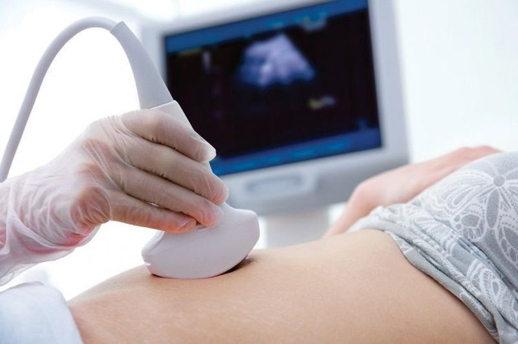
Most fetal abnormalities (68%) identified at 35 to 37 weeks of gestation were diagnosed during the first and/or second trimester, with only about 0.5% being detected for the first time during this period. An ultrasound at this stage includes an examination of fetal anatomy and biometry, assessing fetal growth, fluid volume, and Doppler measurements of the cerebral arteries connecting the uterus, umbilicus, and fetus. This can help predict the development of conditions like preeclampsia and identify fetuses that are small or grow slowly.
Furthermore, ultrasounds during this gestational period can detect new fetal abnormalities not identified in prior examinations. Specific issues include:
- Central Nervous System Abnormalities: Conditions such as ventricular dilation, choroid plexus cysts, and microcephaly.
- Facial Abnormalities: For instance, lacrimal duct obstruction.
- Heart Abnormalities: Among cases of ventricular septal defect, 18.3% (22 out of 120) were first diagnosed in the third trimester, with 4.2% (5 out of 120) identified after birth. Most cases of rhabdomyoma were diagnosed during the third trimester, while some conditions like coarctation of the aorta, pulmonary artery stenosis, and tricuspid valve defects might be detected later or post-birth.
- Abdominal Abnormalities: Such as ovarian cysts, dilated kidneys, and duplex kidneys.
A significant number of fetal abnormalities are first detected during routine ultrasounds between 35-37 weeks of gestation. According to research, routine ultrasounds during this period can aid in identifying fetal abnormalities, potentially improving outcomes by allowing for better preparation for safe delivery and postpartum care.
If an ultrasound indicates potential issues, it’s natural for the pregnant woman to feel worried. However, ultrasound findings can encompass a wide range of possibilities, from serious abnormalities to minor concerns. If an issue is suspected, further specific examinations will be conducted to determine the best course of action and preparation for delivery.
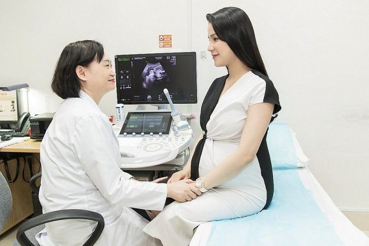
The entire monitoring process is thorough, minimizing the chance of missing any fetal problems, especially those that can be treated before birth or those that require intervention at birth. Vinmec has successfully managed challenging cases such as placenta accreta, umbilical cord knots, and fetal retention. Based on detected abnormalities, doctors will devise an appropriate delivery plan for the pregnant woman.
In addition to a scientific and high-quality examination process, other contributing factors include the modern medical equipment system at Vinmec, which enables rapid intervention for any complications during pregnancy and labor, ensuring the safest possible experience for mothers.
To arrange an appointment, please call HOTLINE or make your reservation directly HERE. You may also download the MyVinmec app to schedule appointments faster and manage your reservations more conveniently.

