This article was professionally reviewed by MD, Specialist level I - Vo Cong Hien - Diagnostic Imaging Department - Vinmec Nha Trang International General Hospital.
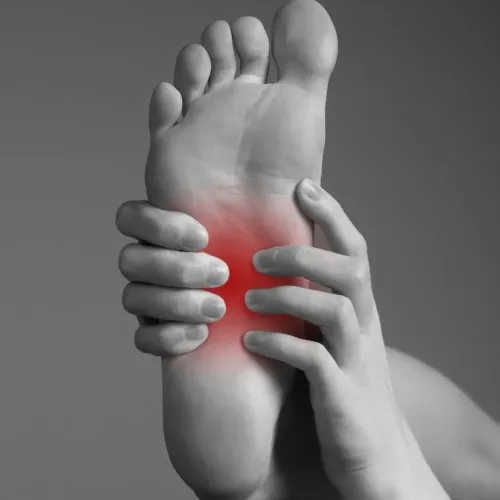
Elastography ultrasound is a technique used to evaluate the mechanical properties of tissues by applying pressure on the probe and detecting tissue displacement through ultrasound waves. This method is currently being researched and applied in diagnosing musculoskeletal disorders.
A rheumatic nodule can be visualized using both conventional (gray-scale) and elastography ultrasound. On a standard ultrasound image, the area appears relatively homogeneous, without clearly defined soft or fluid regions. However, when elastography is applied, softer areas (green, yellow, red) can be visualized, while the surrounding tissues appear stiffer (blue)
1. What is Elastography Ultrasound?
Elastography ultrasound is an imaging technique based on the elasticity and stiffness characteristics of tissues. Different body tissues are composed of various components, giving them distinct stiffness levels. Pathological changes alter tissue stiffness — for instance, cancerous tumors are generally harder than surrounding tissues, and diseased liver tissue is stiffer than healthy portions.
Principle: Under the same applied pressure, soft tissue deforms more, whereas hard tissue deforms less. This deformation is color-coded into an elasticity map.
There are two main types of elastography: Static elastography: tissue response to slow compression. Dynamic elastography: tissue response to rapid compression or vibration.
Another classification includes: Strain elastography (based on tissue deformation) and shear-wave elastography (based on shear wave propagation)

2. Applications of Elastography Ultrasound in Diagnosing Musculoskeletal Disorders
Elastography ultrasound is a new imaging technology based on ultrasound, currently used to assess breast and liver lesions, and is being evaluated for prostate, thyroid, cervical, and lymph node abnormalities. It is also being studied and applied in assessing musculoskeletal injuries.
Numerous studies have demonstrated the effectiveness of elastography ultrasound in diagnosing musculoskeletal conditions, particularly tendon disorders, showing comparable specificity, sensitivity, and accuracy to standard ultrasound in detecting tendon abnormalities in both symptomatic and asymptomatic patients.
Applications in specific musculoskeletal conditions:
- Achilles tendon:
- In healthy individuals: the tendon appears stiff, with dominant blue coloring (indicating hardness and minimal deformation).
- In patients with tendinopathy or achillodynia: the tendon appears soft and homogeneous, predominantly red.
- This distinction is useful for prognosis and detecting subclinical changes before they appear on standard B-mode ultrasound.
- Supraspinatus tendon:
- Frequently injured and often affected by tendinopathy.
- Information about tendon stiffness and elasticity plays a vital role in treatment planning.
- Real-time elastography provides results similar to those of the Achilles tendon: healthy young tendons are stiff (blue), while inflamed tendons are softer (red).
In patients with supraspinatus tears, most of the tendon shows blue and green areas,
- Synovial hypertrophy:
- Common in inflammatory or infectious joint disorders.
- On elastography, infectious synovitis appears soft (red), whereas non-infectious synovitis appears stiffer (blue).
- This distinction is clinically valuable for early management and differential diagnosis in initial stages of synovitis.
- Soft tissue lesions:
- Hemangioma: soft, predominantly red.
- Neurofibroma: firmer, predominantly blue.
- Cysts: typically red, soft, and homogeneous, though sometimes display all three colors due to imaging artifacts.
- Epicondylitis (tennis elbow): swollen and softened common extensor tendon.
- Myositis: elasticity varies — soft in early stages, stiffer (blue) as the disease progresses.
- Neuritis: soft, predominantly red.
Elastography Techniques
Strain elastography: provides quick, semi-quantitative tissue elasticity assessment. It is considered superior to B-mode and Doppler ultrasound for evaluating tendons and monitoring treatment response. Shear-wave elastography: provides more precise quantitative evaluation, overcoming depth limitations.
In summary, studies confirm that elastography ultrasound is a highly promising tool for assessing and diagnosing musculoskeletal disorders.
Elastography ultrasound is a modern imaging technique currently applied to assess a wide range of conditions - including diseases of the breast, hepatobiliary system, prostate, thyroid, cervix, and musculoskeletal system. It is a non-invasive, cost-effective, and highly efficient diagnostic tool.
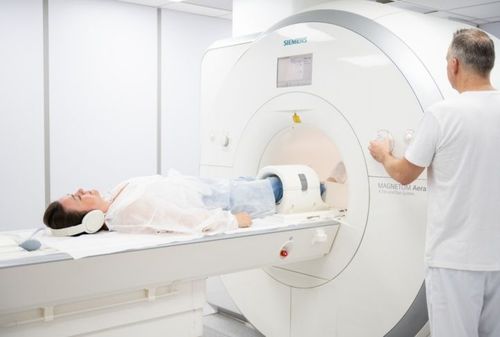
Vinmec International General Hospital is equipped with state-of-the-art diagnostic imaging systems - including MRI, ultrasound, and CT scanners - that meet international standards. Elastography procedures for musculoskeletal and other diseases are performed with precision and adherence to standard protocols by a team of highly qualified physicians using advanced equipment. This ensures accurate results and contributes significantly to disease identification and staging.
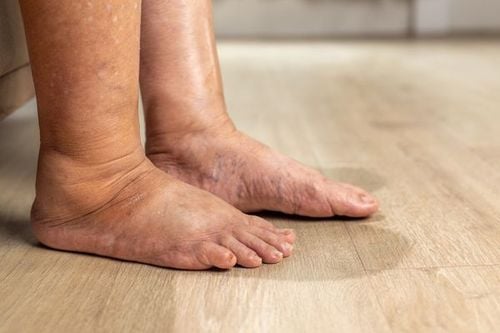
Therefore, patients with musculoskeletal conditions such as rotator cuff tendonitis, elbow tendinopathy, ankle tendon disorders, or other soft tissue lesions should undergo examination and receive timely consultation from specialists.
MD, Specialist level I - Vo Cong Hien, has many years of experience in diagnostic imaging, especially in abdominal ultrasound, advanced cardiovascular ultrasound, and obstetrics-gynecology. Before joining Vinmec Nha Trang, he worked in the Diagnostic Imaging Department at University Medical Center - Hoang Anh Gia Lai.
For consultations or to schedule high-precision diagnostic imaging at Vinmec, please register HERE
Source: ncbi.nlm.nih.gov
To arrange an appointment, please call HOTLINE or make your reservation directly HERE. You may also download the MyVinmec app to schedule appointments faster and manage your reservations more conveniently.