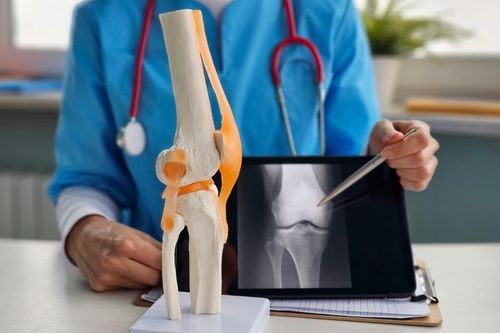
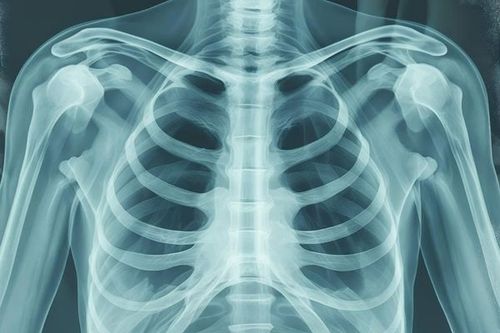
The sternum is a long, flat bone located in the middle of the chest, forming part of the rib cage and connecting the ribs. Tumors beneath the sternum may originate from the sternum itself or from organs in the anterior mediastinum beneath the sternum, such as the thymus gland, connective tissue, and fat.
1. Types of Tumors Beneath the Sternum
1.1. Sternal Tumors
Tumors of the chest wall, particularly those arising from the sternum, are extremely rare, with an average age of diagnosis reported at 42 years. Sternum tumors may be primary, metastatic, or arise from adjacent tissues. Histologically, they can be malignant, benign, or inflammatory. Malignant tumors of the sternum typically present with pain and swelling.
1.2. Thymic Tumors
Thymic tumors are anterior mediastinal tumors located beneath the sternum. They account for about 30% of anterior mediastinal tumors in adults and 15% in children. According to statistics from the American Cancer Society, the incidence of thymic tumors is approximately 15 per 100,000 people, with similar rates in men and women. Thymic carcinoma is the most common histological type. However, all types of thymic tumors have the potential to invade surrounding tissues and are generally considered malignant.
The exact causes and risk factors for thymic tumors remain unclear. Clinically, thymic tumors occur in both children and adults. For thymomas without myasthenia gravis, the most common age range is 50–69 years, while thymomas with myasthenia gravis usually occur between 30 and 69 years.
Symptoms vary widely, from asymptomatic cases detected incidentally on chest X-rays to patients presenting with dry cough, chest pain, dyspnea, or myasthenia gravis. Approximately one-third to one-half of patients with thymic tumors are asymptomatic. Another one-third present local symptoms such as chest pain, cough, or difficulty swallowing, while the remaining third are diagnosed after developing myasthenia gravis (an autoimmune mechanism).
Generalized myasthenia gravis symptoms (muscle weakness, respiratory failure) occur in 70% of cases. The remaining 30% exhibit ocular myasthenia with mild symptoms such as ptosis. Myasthenia gravis can serve as a clinical marker for early thymic tumor detection, leading to earlier diagnosis in affected patients. Rarely, thymic tumors have distant metastases at diagnosis.
Thymic tumors are classified into four stages. Stages I and II often lack obvious clinical signs, whereas stages III and IV manifest symptoms due to tumor invasion and compression of mediastinal structures.
Common compression symptoms include superior vena cava syndrome (facial swelling, chest pain, dyspnea), phrenic nerve palsy causing diaphragmatic paralysis and breathing difficulty, and pleural effusion if the tumor invades the pleura.

2. Diagnosis of Tumors Beneath the Sternum
2.1. Imaging
Various imaging modalities assist diagnosis, each with advantages and limitations:
- X-ray: Limited value in diagnosing sternum or sub-sternal tumors.
- Computed Tomography (CT): The preferred method for evaluating sternum tumors and thymomas. Chest CT accurately determines tumor location, invasion extent into adjacent structures, and detects pleural metastases. It also evaluates other thoracic components.
- Magnetic Resonance Imaging (MRI): Particularly valuable when the tumor invades major vessels. MRI supplements CT by assessing tumor spread beyond the dura and involvement of surrounding structures. It is especially useful in surgical planning.
- Positron Emission Tomography (PET): Currently, PET has no defined role in thymic tumor diagnosis.
2.2. Histopathology
Histological examination confirms tumor type. Image-guided needle biopsy can be used, but open biopsy is considered the best approach for comprehensive histological evaluation.
Needle biopsy is less commonly used in thymoma diagnosis due to lower accuracy. However, combining it with immunohistochemistry improves diagnostic precision. Needle biopsy is mainly reserved for cases where tumors are extensive and require pre-treatment diagnosis before chemotherapy or radiotherapy or when differentiating lymphoma or seminoma—tumors not treated surgically.
2.3. Endoscopy
Mediastinoscopy under general anesthesia allows biopsy with 95–100% accuracy in histological diagnosis.
3. Treatment of Tumors Beneath the Sternum
3.1. Treatment of Sternum Tumors
Similar to other bone tumors, treatment depends on tumor nature, malignancy, and chest wall reconstruction potential.
Sternum cancers often require surgical resection of cancerous cells. Depending on invasion extent, surrounding structures may also need removal.
Surgery typically involves sternum removal followed by chest wall reconstruction. Reconstruction is crucial but challenging to restore chest wall integrity.
Chemotherapy and radiotherapy may be combined with surgery. These treatments can be used preoperatively to shrink tumors or postoperatively to eliminate residual cancer cells.
3.2. Treatment of Thymic Tumors
Treatment involves symptom management (mainly myasthenia gravis treatment) and tumor management.
Patients with myasthenia gravis or suspected cases should be carefully evaluated by neurologists and treated with corticosteroids and cholinesterase inhibitors.
Severe generalized muscle weakness caused by myasthenic crisis requires aggressive treatment such as plasmapheresis or plasma exchange to reduce circulating autoantibodies. These measures help avoid corticosteroid use before surgery, which may impair wound healing. After thymectomy, myasthenia symptoms usually improve, with complete remission rates up to 63%.
Treatment modalities include surgery, chemotherapy, radiotherapy alone, or combined, depending on disease stage. Early-stage thymic tumors have a good prognosis. Surgery is the cornerstone treatment significantly affecting patient survival.
Surgery is indicated when tumors are operable (stages I, II, III, IVA) and without distant metastasis. Most thymic tumors are radiosensitive; thus, radiotherapy is used postoperatively for stage II–IV tumors or after non-radical surgery or open biopsy. Chemotherapy may be used as adjuvant or neoadjuvant therapy for advanced, inoperable thymic tumors.
For patients at stage II or above with myasthenia gravis, neoadjuvant chemotherapy or concurrent chemoradiotherapy followed by surgery reduces complications and mortality, increasing chances for complete resection. However, neoadjuvant treatment may impair precise staging.
To arrange an appointment, please call HOTLINE or make your reservation directly HERE. You may also download the MyVinmec app to schedule appointments faster and manage your reservations more conveniently.