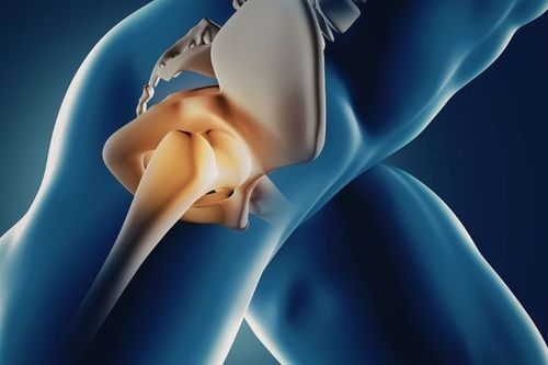

In the human body, the spine is the main pillar that supports the entire body. The vertebrae of the spine, together with the ligaments and intervertebral discs, form a spinal canal that protects the spinal cord inside.
Medical content reviewed by MSc, MD. Nguyen Thi Thanh Binh - General Surgery Department, Vinmec International Hospital Da Nang
1. Length and Shape of the Human Spine
The spine is the central supporting structure of the human body. It keeps the body upright and connects other parts of the skeleton together: the head, chest, pelvis, shoulders, arms, and legs. Although the spine is made up of a series of bones, it is highly flexible due to its system of elastic ligaments and intervertebral discs.
The length of the spine depends on a person’s height. The average length is 71 cm in men and 61 cm in women. The human spine has multiple functions: it bears the weight of the head, trunk, and arms, and allows the body to move in all directions. Some parts of the spine are more flexible than others, with the most flexible part being the cervical spine. The bones forming the spine also protect the spinal cord.
When viewed from the side, the spine has a natural curvature. This S-shaped curve makes the spine stable, helps maintain balance in the upright position, functions as a shock absorber during walking, and protects individual vertebrae from fractures.
2. How Many Vertebrae Does the Human Spine Have?
The human spine is usually made up of a total of 32–34 vertebrae, divided into five regions:
- Cervical segment (7 vertebrae): Labeled from C1 to C7 (C stands for Cervical). This segment has small vertebral bodies with wide transverse diameters. The pedicles are attached to the lateral surfaces of the vertebral bodies rather than the posterior.
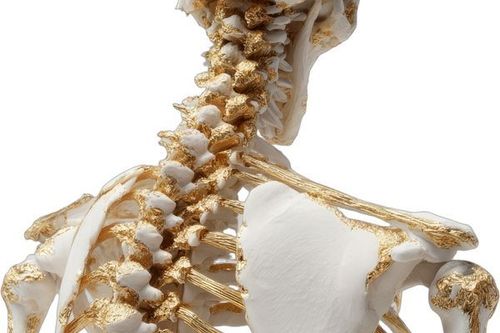
- Thoracic segment (12 vertebrae): Labeled from T1 to T12 (T stands for Thoracic). The spinous processes are long and slanted downward; the transverse processes have articular surfaces that articulate with the heads of the ribs to form costovertebral joints.
- Lumbar segment (5 vertebrae): Labeled from L1 to L5 (L stands for Lumbar). The vertebral bodies are large and wide, with thick pedicles and rectangular spinous processes.
- Sacral segment (5 vertebrae): Labeled from S1 to S5 (S stands for Sacrum). These vertebrae are fused together to form the sacrum, which articulates with the pelvis through the sacroiliac joints.
- Coccygeal segment (3–5 vertebrae): These vertebrae fuse together to form the triangular-shaped coccyx (tailbone).
3. Intervertebral Discs
There are 23–24 elastic intervertebral discs between the vertebrae, except between the skull and the first cervical vertebra, between the first and second cervical vertebrae, and between the sacrum and coccyx, which are fused and do not have discs.
The discs are biconvex and lens-shaped, 3–9 mm thick, highly elastic, composed of cartilaginous and fibrous layers with a tough outer ring. Inside the disc lies a gelatinous nucleus pulposus, spherical or oval in shape, surrounded by concentric layers of fibrous rings. The nucleus pulposus is usually located in the posterior two-thirds of the disc.
The total height of all intervertebral discs accounts for one-fourth of the total height of the spine. They are radiolucent on standard X-rays. The discs maintain spinal flexibility, allowing the body to bend and twist, and they absorb shock from impacts such as running or jumping.
For example:
When force is applied to the spine, the discs are compressed and become thinner. When the pressure is relieved, the discs reabsorb fluid and expand (similar to decompression). Because we put more pressure on the spine during the day and less at night, the spine becomes about 1.5–2 cm shorter by the end of the day.
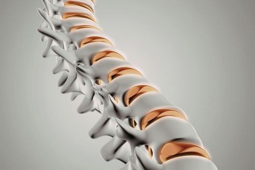
Over the years, as we age, the spinal discs lose elasticity and become thinner, the vertebrae move closer together, the vertebral bodies decrease in height due to reduced bone density, and the spine becomes more curved. This is why people tend to lose a few centimeters in height as they get older.
4. Neural Transmission Within the Spinal Canal
The spinal canal is formed by the stacking of vertebral foramina. The canal is wider in the cervical and lumbar regions than in the thoracic region. The spinal canal is triangular in shape, except in the thoracic region where it is round. The transverse diameter of the canal is determined by the distance between the pedicles on frontal X-rays, and the anteroposterior diameter is measured from the posterior surface of the vertebral body to the anterior surface of the inferior articular process.
Nerve root canals: Except for the first two cervical vertebrae (C1 and C2), the sacrum, and the coccyx, each vertebra consists of a vertebral body anteriorly and bony processes projecting posteriorly. At the junction of two adjacent vertebrae, openings are formed on both the left and right sides - these are the intervertebral foramina, through which spinal nerves exit the spinal canal.
The spinal nerves carry nerve signals from the brain to the skeletal muscles and internal organs through the spinal cord. Similarly, they transmit sensory information such as touch, pressure, temperature, pain, and other sensations from the skin, muscles, joints, and internal organs to the brain via the spinal cord. Together, the spinal cord and brain form the central nervous system.
To arrange an appointment, please call HOTLINE or make your reservation directly HERE. You may also download the MyVinmec app to schedule appointments faster and manage your reservations more conveniently.