The article is professionally consulted by Dr. Nguyễn Anh Tú - Specialist of Obstetric ultrasound & prenatal diagnosis - Department of Obstetrics - Vinmec Hai Phong International General Hospital.
The journey of fetal development within the womb is truly remarkable. Mothers often wonder how their baby is growing and developing each week. This article provides an in-depth look at the formation and weekly development of the fetus.
1. Stage 1: Embryonic Formation
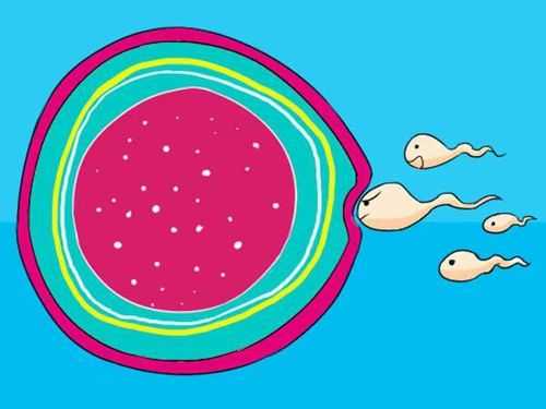
- Milestone 1: As the vaginal canal expands, it creates a highly acidic environment, over 60 million sperm are released into the vagina, with only a fraction making it to the cervix.
- Milestone 2: The cervix contains a thick mucus layer, making it difficult for sperm to pass through. Only about 100,000 sperm successfully navigate through this barrier.
- Milestone 3: Sperm move at a speed of 5 mm per minute, reaching the cervix in just 10 minutes. However, white blood cells detect the presence of foreign cells and destroy many sperm. Ultimately, only 200 sperm manage to reach the fallopian tubes.
- Milestone 4: The fallopian tubes measure approximately 10 cm, and the sperm must complete this final journey to reach the mature egg. Only the strongest sperm will succeed in penetrating the egg's protective barrier.
Once the selected sperm reaches the egg, it releases enzymes that help it breach the outer membrane. Meanwhile, the egg seals off all entry points. The fusion of sperm and egg forms a zygote.
2. Stage 2: Fetal Development by Week
- 1-Week-Old Embryo: At this stage, the uterine lining thickens to support and nourish the fertilized egg. If fertilization does not occur, this lining will shed during the next menstrual cycle.
- 2-Week-Old Embryo: Fertilization typically takes place in the fallopian tube. However, at this point, the mother will not notice any visible signs of pregnancy.
2.1. First Trimester
The first 3 months of pregnancy mark significant physical and hormonal changes in the mother’s body. During this phase, fetal growth is gradual, as the baby is establishing a foundation for further development.
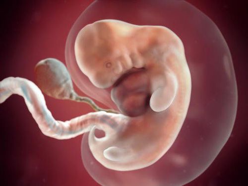
- 3-Week-Old Embryo: This is the time that can confirm successful conception. The first noticeable sign is a missed period. Fertilized egg, or zygote, begins its journey into the uterus, searching for the optimal implantation site for the upcoming nine months. The mother’s body responds by increasing the production of estrogen and progesterone, two key hormones responsible for sustaining pregnancy.
- 4-Week-Old Embryo: Early pregnancy symptoms may still be undetectable for some mothers. The fertilized egg implants itself into the uterine lining, forming a blastocyst, a ball-like structure consisting of hundreds of cells that will later develop into the fetus. At this stage, the embryo starts producing hCG (human chorionic gonadotropin), the pregnancy hormone that prevents monthly ovulation. Furthermore, the embryo will begin cellular activity to form the initial structural framework of the fetal body.
- 5-Week-Old Embryo: By 5th week, the embryo is officially formed. It has been 4 weeks since the mother’s last menstrual period. During this week, early pregnancy symptoms start becoming apparent. The 5-week-old embryo is microscopically small but has already grown 10,000 times larger than the embryo at fertilization. Moreover, during this time, a pregnancy test can now confirm the pregnancy.
- 6-Week-Old Embryo: This is the ideal time for the first prenatal check-up. By 6th week, the embryo can be measured via ultrasound, with an average gestational sac length of 5–6 mm. The fetus is now approximately 1 cm in length, similar in size to an apple seed, and resembles a tadpole-like structure. During this period, rapid development occurs, with the formation of the circulatory system and skeletal framework. Most significantly, the fetal heart begins to beat.
- 7-Week-Old Embryo: By 7th week, the fetal heartbeat can be clearly detected via ultrasound, which is the first sign showing that the baby is growing inside the mother's body. The fetus is now the size of a pea. During this period, the baby’s brain is undergoing rapid development. Additionally, the liver has started producing red blood cells, a function that will later be taken over by the bone marrow. Facial features, including the nose, mouth, and ears, also begin to take shape. This is also an important milestone when many mothers start experiencing morning sickness.
- 8-Week-Old Embryo: Your baby is now officially called a fetus, measuring approximately 1.5 cm in length. At twice the size of week 7, the fetus still has a small tail. However, this tail will soon disappear. The limbs are paddle-shaped but are beginning to develop more distinct arms and legs. The heart valves and air passages connecting the throat to the lungs are now forming. Fingers, toes, lips, and eyelids are becoming more defined. By week 8, your baby is about the size of a blueberry.
- 9-Week-Old Fetus: At 9th week, the fetus has grown to 2.3 cm, approximately the size of a grape. The reproductive organs are beginning to form, but the gender cannot be accurately determined until weeks 15-17. The mouth and tongue are developing, and the hands are now divided into individual fingers. Although the baby has started to move, these movements are still too subtle for the mother to feel. The neurons are branching out and forming the first neural pathways.
- 10-Week-Old Fetus:The heart has fully developed into four chambers and the fetal heartbeat can be detected using Doppler ultrasound. While all major organs and body structures are already developed, the brain remains disproportionately large, and the digestive system is still developing. During this period, the development physiology is basically complete, even tiny earlobes are forming, but many body systems will continue to mature in the coming weeks. The tail from early embryonic development has now disappeared, and the fetus is about the size of a cherry.
- 11 Week-Old Fetus: The baby’s larynx is forming, though it will take more time to fully develop. The hands can now move and form a fist. Meanwhile, the brain and nervous system continue to grow rapidly. Although the genital structures have formed, it is still difficult to determine the gender. This week, the tooth buds emerge, eyes are fully developed, fine hair begins to cover the body and the baby's facial features become more distinct. At this time, the fetus is now the size of a strawberry.
- 12 Week-Old Fetus: The baby’s body appears more structured and well-formed, resembling a newborn more than ever before. The central nervous system, heart, liver, and excretory system have developed their basic functions. The 12th week is also a crucial milestone for prenatal ultrasound, allowing doctors to monitor fetal heart rate, which should be between 120 - 160 beats per minute in a healthy pregnancy. At the 12th week, the baby is about the size of a lemon.
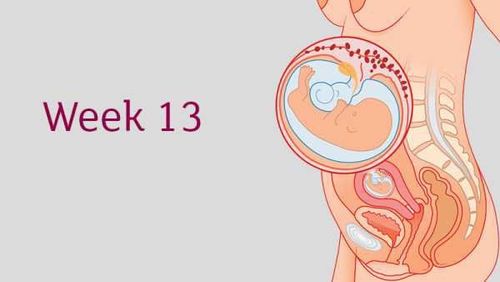
- 13 Week-Old Fetus: At week 13, the baby begins involuntary reflex movements, such as opening and closing fingers, curling toes, and making sucking motions with the mouth. However, these movements are not yet strong enough for the mother to feel. The baby is now the size of a plum and can respond to external stimuli, such as light touches on the belly. By this stage, the unique fingerprint pattern has already formed. Moreover, the baby can even frown or make facial expressions inside the womb.
2.2. Second Trimester (Weeks 14–27)
The second trimester, from week 14 to 27, is considered the most stable phase of fetal development. During these 3 months, the fetus experiences significant growth in bones and muscles, and ultrasound is able to provide clearer facial features of the baby.
- 14 Week-Old Fetus: From week 14 onward, fetal size and weight increase significantly, with an average weight gain of 2 grams per week. The central nervous system continues to develop, reaching millions of nerve cells, while the reproductive organs become more defined.
- 15 Week-Old Fetus: At week 15, the fetus measures approximately 10.1 cm in length and weighs around 70 grams, about the size of a small apple. Although the eyelids remain closed, the fetus can perceive light passing through the mother’s abdomen. The baby starts to have brain activity and facial movements. The kidneys now become functional. Ultrasound may capture thumb-sucking behaviors. By this week, the fetus is the size of a peach.
- 16 Week-Old Fetus: By week 16, the bones have strengthened, making the fetus more robust. This is also an ideal time for pregnant women to maintain adequate calcium intake to support bone development as well as to engage in prenatal education techniques to stimulate brain development.
- 17 Week-Old Pregnant: At this stage, the fetus can now move its joints, and sweat glands begin to develop. The fetus measures approximately 13 cm in crown–rump length and weighs about 140 grams, similar in size to an avocado. The scalp structure is forming, but hair is not yet visible. The legs have grown longer, and the baby’s kicks may now be felt. The head is straighter, and the ears are gradually moving into their final position.
- 18 Week-Old Fetus: The fetus now appears more proportionate, with limbs developing symmetrically. A few strands of hair are starting to emerge on the head. By week 18, the baby is the size of a pomegranate and can move its joints freely. The skeletal system, initially composed of soft cartilage, is beginning to ossify. The umbilical cord has also become thicker and stronger.
- 19 Week-Old Fetus: The mother may clearly feel the baby's heartbeat from within. The reproductive organs are now fully formed, allowing gender determination via ultrasound. The fetus is now the size of an artichoke. He/she now can bend its legs, and these movements may be noticeable. Meanwhile, myelin sheaths are starting to form around developing nerve fibers.
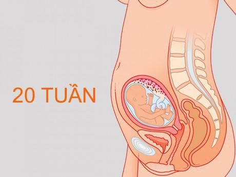
- 20 Week-Old Fetus: By week 20, the fetus experiences a significant growth spurt, reaching 16.4 cm in crown–rump length and weighing around 300 grams. The baby actively swallows amniotic fluid, preparing for postnatal digestion. Although the eyelids remain closed, pupillary movements may begin. The five sensory systems such as smell, sight, touch, taste, and hearing, are rapidly developing, and the fetus can now hear external sounds, including the mother’s voice. At this stage, the baby is the size of a mango.
- 21 Week-Old Fetus: This week marks the formation of the fetal jawbone, while the arm and leg muscles become stronger, preparing the baby for the quickening stage. By week 21, hair and eyelashes begin to grow. The baby can now swallow amniotic fluid, and the digestive system starts producing meconium, the thick, dark stool that will be passed after birth. The fetus is now the size of a banana.
- 22 Week-Old Fetus: By week 22, the fetus resembles a newborn in structure. Weighing around 430 grams and measuring 26.7 cm from head to bottom, the baby is now the size of a small pumpkin. Movements become more forceful, sometimes causing sharp sensations in the mother’s abdomen.
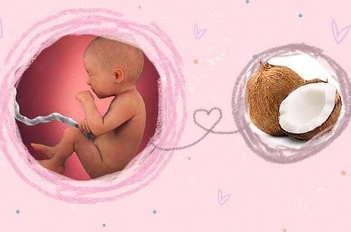
- 23 Week-Old Fetus: The baby’s nostrils are now open. Facial features are well-defined, and the body is becoming rounder. At this stage, the baby resembles a newborn but remains small, about the size of a coconut. However, the eye pigmentation has not developed yet.
- 24 Week-Old Fetus: This time marks the halfway through pregnancy. During this stage, the baby begins accumulating fat deposits under the fingers, palms, and soles. The skin stretches to accommodate increasing fat storage. The nervous system develops rapidly, the eyes begin to blink, and the ears can now recognize the voices of both parents. The sensation becomes more sensitive, enhancing the baby's ability to perceive external stimuli. A particularly exciting milestone for mothers is the first noticeable fetal movements. Additionally, the first fat deposits start accumulating under the fingers, palms, and soles, helping the skin stretch and prepare for further fat storage until birth. At this stage, the baby is about the size of a grapefruit.
- 25 Week-Old Fetus: The baby’s skin remains thin and translucent, allowing blood vessels to be visible on 3D and 4D ultrasound. The fetus weighs approximately 660 grams and head-to-bottom length measures 34.6 cm. Though appearing long and slender, muscle mass and fat deposits are increasing. The skin is still delicate and transparent but will become more thickening soon. The baby is now the size of a cantaloupe.
- 26 Week-Old Fetus: The baby now sleeps in short cycles to support brain and visual development. By week 26, the fetus is the size of a cauliflower, with wrinkled skin that will smooth out as fat accumulates. This helps your baby look more like a newborn. Hair begins to grow, and its color and texture become more defined. The baby weighs around 900 grams, and the head is now proportionate to the body. During sleep, significant brain development occurs.
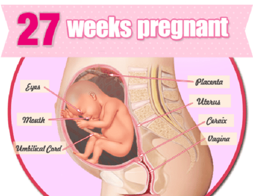
- 27 Week-Old Fetus: By week 27, the baby gains an additional 300 - 400 grams. The baby spends most of the time sleeping to conserve energy and store necessary fat. The baby is now the size of a kale leaf. He/she also practices breathing by inhaling and exhaling amniotic fluid, an essential preparation for life outside the womb. This is also the most active phase of fetal movement, which indicates healthy development.
2.3. Third Trimester
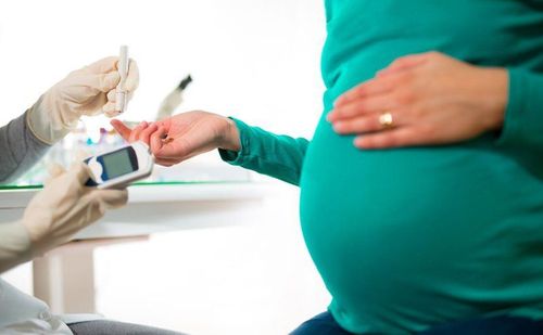
During the final 3 months of pregnancy, the fetus primarily focuses on weight gain and completing organ development in preparation for birth. Pregnant women should be aware of potential complications such as preterm labor, gestational diabetes, or preeclampsia, which may arise if maternal health becomes unstable.
- 28 Week-Old Fetus: The baby’s brain grows rapidly, making it essential for pregnant mothers to consume foods that are good for brain development, such as fish. By now, the fetus weighs around 1 kg, and its head size adjusts to accommodate advanced brain development. The baby has strong arms and legs and continues to swallow amniotic fluid while absorbing fat to regulate body temperature after birth. At this stage, the baby is the size of a Napa cabbage.
- 29 Week-Old Fetus: At week 29, vision continues to develop, and the baby can detect external light. He/she also can blink, and eyelashes continue to grow. The current visual acuity is 1/20, which will improve over time. The baby also begins recognizing parental voices, making regular conversations with the baby beneficial.
- 30 Week-Old Fetus: The baby can now open and close its eyes frequently. Moreover, the head grows larger to accommodate brain expansion. Lung and muscle activity increase, preparing for postnatal functions. During this stage, the baby still sleeps most of the time. However, fetal movements are clearly visible during his/her awake time, such as knee jabs, elbow pushes, and rhythmic hiccups.
- 31 Week-Old Fetus: By week 31, the lungs are fully developed, which means the baby can breathe independently if born prematurely. The baby is the size of a young coconut, measuring 41.2 cm in length and is covered in 1.5 liters of amniotic fluid. As the baby grows larger, amniotic fluid volume will gradually decrease. The baby gains weight rapidly in preparation for birth.
- 32 Weeks Pregnant: This week, the baby begins shifting into a head-down position in preparation for birth. From this stage, there will be no significant changes in the baby’s weight. Fat deposits accumulate under the skin, making the baby plumper. While movement patterns become more regular, intentional and goal-directed movements will only develop in the coming months. At this time, the movement and resting of the fetus follow a fairly regular cycle.
- 33 Week-Old Fetus: At this stage, the baby weighs 2 kg with head to bottom length at 43.7 cm. He/she now also has a more stable body temperature, requiring less dependence on the mother’s warmth. The brain continues to grow rapidly, making DHA-rich foods essential for development. By week 33, the baby is the size of a squash.
- 34 Week-Old Fetus: The baby now produces meconium inside the intestines. Bones are almost fully developed, except for the skull, which remains soft and flexible to facilitate passage through the birth canal. This week, the skull bones have not yet fused, allowing for easier vaginal delivery. Mothers may feel relief from breathing difficulties as the baby shifts downward but may experience increased bladder pressure. The baby is the size of a large celery stalk.
- 35 Week-Old Fetus: This week, the baby is 2.3 kg and 46.2 cm. The baby’s organs are fully functional. If born this week, the baby is likely to thrive without complications. The central nervous system and lungs continue their final maturation. Babies born between weeks 34-37 are generally healthy and capable of independent breathing.
- 36 Week-Old Fetus: By week 36, the baby weighs 2.6 kg and measures 47 cm. Now, the baby is roughly the size of a pineapple, which makes it feel tighter in the uterus. The kidneys and liver are fully developed, capable of excreting waste products. Most organ systems are complete, except for the brain and lungs, which will continue developing after birth.
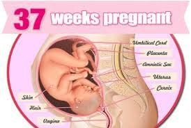
- 37 Week-Old Fetus: The baby is now head-down, continuously pressing against the mother’s pelvis. Weight gain averages 30 grams per day. A protective white coating (vernix caseosa) forms on the skin, preventing dryness. By week 37, the baby is the size of a papaya. If born this week, it is considered late preterm birth, with minimal health risks.
- 38-week-old fetus: By week 38, the delivery date is approaching, and although the baby now closely resembles a newborn, it is still not fully ready to enter the outside world. Over the next two weeks, the lungs and brain will continue to fully mature. At 38 weeks, the fetus is considered to be in the final stage of a full-term pregnancy. The subcutaneous fat layer will continue to thicken slightly, helping the baby maintain body temperature after birth.
- 39-week-old fetus: At 39 weeks, the baby engages in essential reflex activities, such as sucking, breathing, swallowing, digesting, excreting, and crying, all of which will continue after birth. While the baby is considered full-term, in some cases, it may not yet be ready for delivery and will remain in the womb for a bit longer.
- 40-week-old fetus: By week 40, the baby's physical development is complete, but fat accumulation continues, helping regulate body temperature after birth. At this stage, the baby is the size of a large pumpkin.
3. Stage 3: Welcoming the Baby into the World
Upon delivery, the baby remains connected to the mother via the umbilical cord, which measures approximately 50 cm in length. This cord has served as the primary lifeline, delivering oxygen and nutrients from the mother while also removing fetal waste products through the maternal excretory system.
After delivery, the umbilical cord is cut, leaving a 2–3 cm stump attached to the baby’s abdomen. Over time, this stump dries and naturally falls off, forming the baby’s permanent navel. In many hospitals today, laser cord-cutting techniques are utilized immediately after birth to enhance safety and reduce the risk of infection.
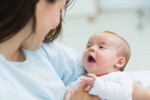
The first week after birth, the so-called perinatal period, is crucial, as the baby must adapt to a new environment outside the womb. The first month, or neonatal period, is considered the most vulnerable phase of life, with the highest risk of mortality due to potential complications.
The 9-month, 10-day pregnancy journey is an incredible experience, filled with surprises and milestones. At Vinmec International Hospital, comprehensive maternity care packages are available, offering exceptional quality and continuous support from experienced obstetricians throughout pregnancy, labor, and postpartum recovery.
Vinmec maternity care packages are listed as following:
- 12-Week Comprehensive Maternity Package
- 27-Week Comprehensive Maternity Package
- 36-Week Comprehensive Maternity Package
- Labor & Delivery Package
Dr. Nguyễn Anh Tú has 6 years of experience in obstetric ultrasound and prenatal diagnostics, with advanced training in fetal ultrasound and prenatal screening. He has completed certified courses from the Fetal Medicine Foundation (FMF) and has received specialized training in prenatal diagnostic and interventional techniques. Additionally, Dr. Tú has participated in numerous international conferences and workshops on fetal medicine. He is currently a specialist at the Department of Obstetrics and Gynecology, Vinmec Hai Phong International Hospital.
To arrange an appointment, please call HOTLINE or make your reservation directly HERE. You may also download the MyVinmec app to schedule appointments faster and manage your reservations more conveniently.