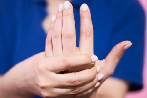
The process of treating phalangeal fractures mostly uses simple treatment methods. In contrast, surgery will be applied in cases of severe finger fractures. However, patients need to receive timely emergency care to avoid dangerous complications that may affect their health.
This article is written under the professional guidance of doctors from the Department of Orthopedic Trauma & Sports Medicine, Vinmec International General Hospital.
1. Phalangeal fractures
1.1. General structure
A very common hand injury is a phalangeal fracture (also known as a phalanx fracture).
Normally, to perform specific tasks such as grasping or manipulating objects, the bones of the hand are perfectly aligned. However, when a finger fracture occurs, this alignment is disrupted, leading to the hand’s inability to perform normal functions.
In fact, this type of bone is one of the most frequently fractured bones in the body, accounting for nearly 10% of all fracture cases.
For young individuals under 30 years old, sports activities are often the cause of finger fractures. Meanwhile, for those aged 30 to 70, occupational and machinery-related accidents are the main causes of finger fractures. In the elderly over 70, falls are usually the reason for finger fractures.
Finger or fingertip fractures, especially when the wound is “closed,” are easily mistaken for other injuries such as sprains or severe dislocations. Therefore, patients should immediately see a doctor for timely medical care.
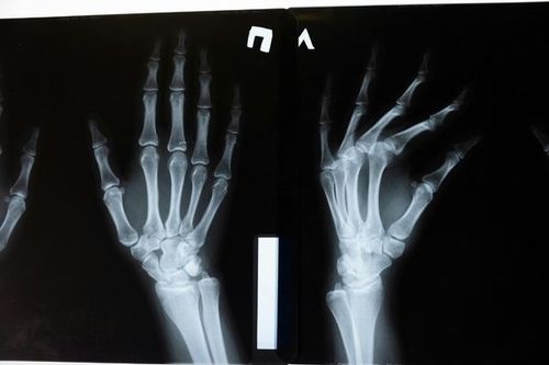
Not only does it help determine the extent of injury, but an X-ray of the affected finger also allows doctors to know whether the finger or fingertip is fractured or not.
Anatomically: Each hand consists of 27 bones, including 8 carpal bones, 5 metacarpal bones, and 14 phalanges. Each finger has 3 phalanges: the proximal phalanx is the first bone of the finger, the middle phalanx is the middle bone, and the distal phalanx is the fingertip bone.
In this section, proximal and middle phalanx fractures are the main focus.
1.2. Causes of finger fractures
Among the parts of the hand, the fingers are the most vulnerable. Finger injuries can vary greatly, from bruises due to impact to infections or fractures. In addition, finger injuries and fractures are also common conditions.
Compared to other parts of the body, fingers are more prone to injury simply because the hands and fingers are used most frequently. Some different ways that people may injure their fingers or fingertips include:
- Using saws or hammers at work.
- When contacting fast-moving objects, including sports equipment such as cricket balls, fingers are more likely to fracture.
- Finger or fingertip fractures can occur when a person accidentally slams their hand in a car door.
- When trying to break a fall or slip by extending the hands, a person can fracture a finger.
1.3. Types of finger fractures
- Finger fracture: The ligament or tendon attached to the finger bone is pulled out of its original position.
- Impact fracture: The fractured parts of the finger bone remain aligned.
- Split fracture: Due to applied force, the finger bone splits into two, and each part moves in a different direction.
- Open fracture: The finger bone breaks and pierces through the skin.
- Closed fracture: The bone is not visible through the intact skin.
- Non-displaced fracture: Although fractured, the bone remains in its original position without displacement.
- Comminuted fracture: The finger bone breaks into multiple misaligned pieces.
- Traumatic fracture: An unstable fracture where the bone breaks into three or more fragments
1.4. Symptoms
To obtain an accurate diagnosis and timely treatment of phalangeal fractures, patients should see a doctor as soon as possible. The symptoms of finger fracture include:
- The finger is painful, swollen, and bruised.
- The affected finger becomes stiff and difficult to move.
- A sign that a finger is fractured or dislocated is when it points in an abnormal direction.
- The finger loses sensation and becomes numb.
- The patient can see the bone or the bone protruding through the skin.
1.5. Diagnosis
When a patient reports finger injury, pain, and swelling, the first diagnostic consideration is usually a finger fracture.
A clear sign indicating a finger fracture, dislocation, or both is when the finger is bent abnormally out of its normal position. In addition, the inability to bend the finger due to pain is another sign of this condition.
X-ray imaging is the next step the doctor will perform if a finger fracture is suspected. Finger X-ray is a good, even the only necessary test to diagnose this condition. After that, the X-ray results will be used to guide the treatment of the phalangeal fracture.
2. Treatment of phalangeal fractures
When a person is found to have a finger fracture, the first action the first responder should take is to bring the injured person to the nearest emergency room for immediate care. The reason is that the wound may become infected due to environmental contamination, which can slow bone healing or even require amputation of the injured finger if infection or delayed emergency care occurs.
2.1. Use of antibiotics and tetanus prophylaxis to prevent infection
Patients will receive a tetanus shot immediately and be given early antibiotic therapy to prevent or treat infection that may have already occurred at the wound site upon admission to the emergency room. The type of antibiotic will be chosen based on the severity of the condition.
2.2. Examination
To assess internal structures and the extent of tissue and bone damage, the doctor will perform X-rays or MRI of the fingers.
Depending on the results, the doctor will gently realign the bones as much as possible to prevent free bone fragments from further damaging soft tissue. Afterward, to limit movement and prevent further injury to the finger bones, the finger will be splinted.
To prevent further tissue damage, stabilizing the fractured bone is extremely necessary. The patient will be prepared for bone fixation surgery. The fracture will be stabilized externally or internally, depending on each specific case.
Internal fixation is usually applied in mild finger fractures with minimal contamination and non-severe injuries. In contrast, external fixation is indicated for more complex and severe finger fractures with high risk of infection and damage.
2.3. Wound cleaning
All dirt, foreign bodies, and unclean skin or soft tissues will be completely removed by the surgeon during the wound irrigation and disinfection process to control infection.
In addition, the surgeon will also clean the bone by removing all debris, foreign objects, and small bone fragments. Missing bone segments will be reconstructed in later surgeries. Once irrigation is complete, the wound will be rinsed with saline.
To prevent further tissue damage, stabilizing the fractured bone is extremely necessary. The patient will be prepared for bone fixation surgery. The fracture will be stabilized externally or internally, depending on each specific case.
Internal fixation is usually applied in mild finger fractures with minimal contamination and non-severe injuries. In contrast, external fixation is indicated for more complex and severe finger fractures with high risk of infection and damage.
2.4. Internal fixation for the treatment of finger fractures
First, the broken bone fragments are realigned to their correct position, then securely fixed with screws and plates placed on the bone surface. Thanks to this method of treating phalangeal fractures, the bone healing process occurs almost naturally.
Surgery to fix the fracture may require some time before being performed because finger fractures may cause significant tissue damage and accompany other injuries.
The surgery aims to restore the fractured bone to its original position and maintain alignment until complete healing.
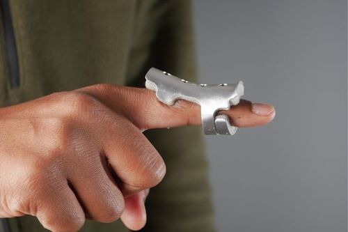
To keep the bone fragments aligned, doctors often use small screws, a small metal plate, or two pins. These metal devices help strengthen bone stability during healing.
Moreover, to ensure bone alignment, multiple X-ray images will be taken during the surgical treatment of phalangeal fractures. If screws are used, they will remain permanently inside the bone, while pins will be removed after 3–4 weeks.
A major advantage of surgery is that patients can begin moving their fingers almost immediately, thereby reducing the risk of joint stiffness.
Additionally, in some cases, fracture-dislocation injuries occur when the fracture extends into the finger joint, causing instability. As a result, the finger becomes hyperextended backward, and both the first and second bones are damaged: the first bone is fractured, and the second bone is dislocated (meaning the finger joint is separated).
The immediate goal is to reposition the finger to its original alignment, then maintain it in that position during healing. When the fracture fragment occupies less than 40% of the joint surface, splinting the finger in a flexed position (with an extension block splint) will allow the bone to heal. Conversely, if the fracture fragment involves more than 40% of the joint, surgery is necessary to restore joint stability.
2.5. External fixation for the treatment of finger fractures
In this method of treating phalangeal fractures, the broken bone is restored to its original anatomical position. Next, screws and plates are fixed above and below the fracture site. Finally, these screws are connected to metal rods placed outside the skin.

After fixation of the compound fracture of the finger bone, doctors will proceed to close the wound. The wound closure method will be chosen depending on the complexity of the wound, using local or free flap techniques.
In the local flap method, damaged wrist muscle tissue is rotated to cover the fracture site, after which a skin graft is applied to the area. In the free flap technique, the entire tissue is completely transferred, usually harvested from the abdominal skin.
For cases of extra-articular finger fractures (away from the joint), the common treatment method for phalangeal fractures is to lightly tape the injured finger to the adjacent finger for about three weeks. If immobilization lasts longer, the risk of finger stiffness increases.
As long as the fractured finger bone remains in normal or near-normal alignment, buddy taping works well. When the bone is fractured at only one site, bent less than 10 degrees, and neither shortened nor rotated, the bone will remain stable.
Because the adjacent finger is used as a splint to protect and stabilize the injured finger while allowing slight movement to prevent stiffness (stiffness is a major concern in finger injuries), buddy taping is very effective.
However, surgery for phalangeal fractures will be indicated when the finger is broken into many small fragments, the fracture extends into the finger joint (intra-articular), or when the bone fragments are excessively displaced from their normal position.
If the finger bone is too short (less than 2 mm) or excessively bent (over 10 degrees), the patient will not be able to fully straighten the finger (due to altered biomechanics).
2.6. Postoperative treatment of finger fractures
After surgery, patients will often feel pain and joint stiffness during the postoperative recovery period for phalangeal fracture treatment. To reduce these symptoms, patients should use nonsteroidal anti-inflammatory pain relievers (NSAIDs) such as Tylenol or anti-inflammatory ibuprofen in combination with gentle exercises.
In addition, to help the fingers return to normal movement early, patients will also be introduced by their doctor to some finger physiotherapy methods.
Reference source: bonetalks.com
To arrange an appointment, please call HOTLINE or make your reservation directly HERE. You may also download the MyVinmec app to schedule appointments faster and manage your reservations more conveniently.